WHAT DOES KNEE OSTEOARTHRITIS LOOK LIKE ON X-RAY?
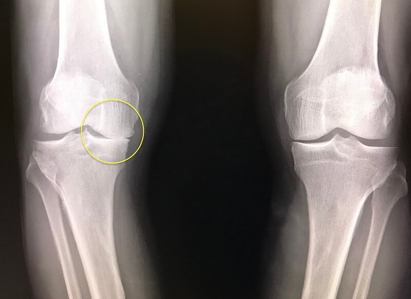
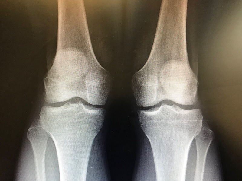
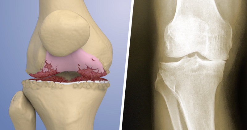
X-rays are extremely helpful in diagnosing the degenerative joint disease that causes OA pain. The main x-ray finding that can confirm osteoarthritis is loss of joint space because of damaged cartilage. As degenerative joint disease gets worse, there is progressive narrowing between the bones because of decreased cushioning from cartilage loss. The amount of joint space loss provides clues to suggest if the osteoarthritis is mild or severe:
+ Mild (less than 50 percent loss of joint space)
++ Moderate (50-90 percent loss of joint space)
+++ Severe (90+ percent loss of joint space/”bone-on-bone”)
There are other x-ray findings that support a diagnosis of osteoarthritis, such as bone spurs and cysts. If x-rays are normal, your doctor may order other tests. An MRI can help identify other injuries in the knee, and can also detect early stages of osteoarthritis before they become visible on your x-rays.
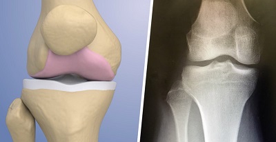
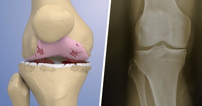
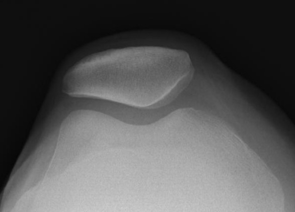
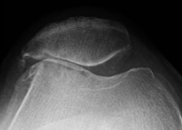
Merchant view x-rays show the patellofemoral (kneecap) joint, where osteoarthritis can occur when cartilage between the bones is damaged and joint space is lost (right).


